Cell Dissociation Protocol using Trypsin
Various proteolytic enzymes are used to detach cells from the adherent substrate, of which the trypsin a member of serine protease family is most frequently used. Trypsin is produced from proenzyme, trypsinogen secreted by exocrine cells of pancreas; Trypsin acts on C-terminal side of Lysine or Arginine. Optimum activity is achieved at 37 °C, so pre-warmed trypsin speed up the detachment. Long term incubation with high trypsin concentration damage cells by striping cell surface proteins and kill the cells.
Trypsin is tolerated by many cell types; however it is desirable to avoid trypsin in proteomic studies and serum-free cultures. Based on the application and cell types trypsin is employed with various constituents and concentration. Some of the attributes are discussed below.
- EDTA (disodium ethylenediaminetetraacetic acid): Adhesion molecules in the presence of calcium determine cell-cell and cell-matrix interaction. To weaken the interactions, EDTA is frequently included with Trypsin which chelates the divalent cations (Ca2+, Mg 2+).
- Phenol red: Optimum activity of trypsin is achieved at pH range from 7 to 9 (5466615). At this range inclusion of phenol red gives pink color. Due to environmental conditions the pH of trypsin may turn acidic, giving orange color and renders trypsin less effective. By adjusting the pH to 7.4 – 7.6 with NaOH trypsin activity could be optimized.
- Diluent: Concentrated trypsin is being solubilized or diluted in a buffered salt solution that contains no Ca2+ or Mg2+, such as Hank's Balanced Salt Solution (Product No. H9394), which maintains pH and osmotic balance. Besides, some trypsin products have 0.9% NaCL as diluent instead of hanks balanced salt solution.
- Source and form: The major source of trypsin is porcine, which is available either in the form of lyophilized powder or as in solution. To avoid animal or microbial products, recombinant bovine trypsin is also expressed in corn, called Trypzean solution (T3449).
- Concentration: Based on the cell type and application trypsin is used in various concentrations. For strongly adherent cell lines, trypsin of 2.5 % to 0.25% (10X to 1X power) is used. While the studies which require cell surface protein integrity, lower concentrated (0.05% trypsin) solutions are employed.
Protocol
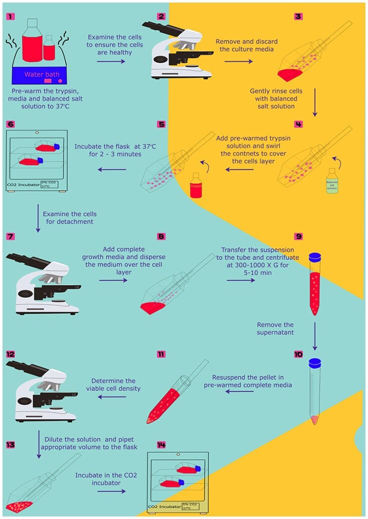
- Pre-warm the trypsin solution, balanced salt solution (Ca+2 and Mg+2-free solution) and growth medium to 37 °C
- Examine the cells to ensure the cells are healthy and free of contamination
- Remove and discard the culture media from flask
- Gently rinse the cells with balanced salt solution without Ca+2 and Mg+2 ions and remove the solution. Firmly adherent cells could also be washed with tryspin solution. However, for sensitive cells balanced salt solution is preferred.
- Add appropriate quantity (0.5 mL/10 cm2) of pre-warmed trypsin solution to the side wall of the flask. Gently swirl the contents to cover the cell layer.
- Incubate the vessel in room temperate for 2-3 minutes. Firmly adherent cells can be detached quickly at 37 °C. Observe the cells under microscope. The detached cells appear rounded and refractile under microscope. If less than 90% of cells are detached incubate the flask for another 2 minutes and observe the cells under microscope for every 30 seconds.
Note: Prevent cell exposure to trypsin solution for longer periods (>=10 min)
- Once cells appear detached add two volumes of pre-warmed complete growth media to inactivate trypsin. Gently disperse the medium by pippeting over the cell layer surface several times to ensure recovery of >95% of cells. For serum free cultures, Soybean trypsin inhibitor (Product No. T6414) is added at an equimolar concentration to inhibit the trypsin.
Note: Vigorous pipetting cause cell damage.
- Transfer the cell suspension to the tube and gently centrifuge at 300-1000 X g for 5-10 min. After removing the supernatant, gently resuspend the cell pellet in pre-warmed complete growth medium. Remove required sample to determine the cell density of viable cells by using hemocytometer and trypan blue exclusion or automated cell counter.
- Dilute the remaining solution to the seeding density and pipet appropriate volume to the culture flask and place it in the incubator.

Figure 1.
For more information, including various other applications and protocols of Trypsin, please click here.
如要继续阅读,请登录或创建帐户。
暂无帐户?