Merck
CN
All Photos(2)

Documents

MAB3412

Sigma-Aldrich

Anti-Cytokeratin AE1/AE3 Antibody, recognizes acidic & basic cytokeratins, clone AE1/AE3

clone AE1/AE3, Chemicon®, from mouse

Sign Into View Organizational & Contract Pricing

Synonym(s):
Pan cytokeratin
eCl@ss:
32160702
NACRES:
NA.41

biological source

mouse

Quality Level

antibody form

purified antibody

antibody product type

primary antibodies

clone

AE1/AE3, monoclonal

species reactivity

chicken, mouse, monkey, human, rabbit, bovine, rat

manufacturer/tradename

Chemicon®

technique(s)

ELISA: suitable
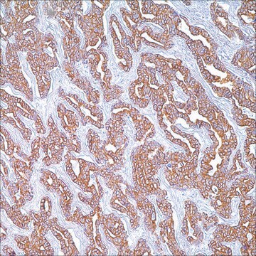
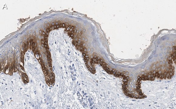
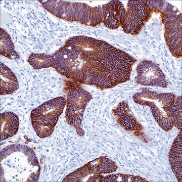
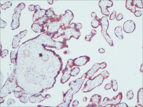
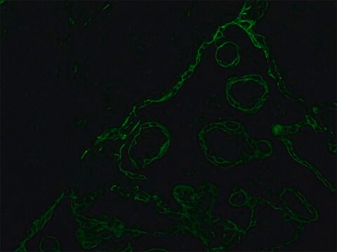
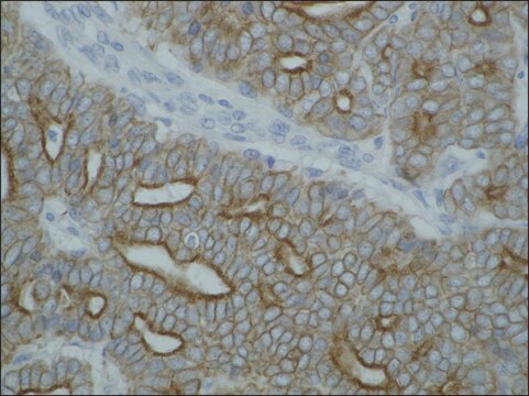
immunohistochemistry: suitable
western blot: suitable

isotype

IgG1

shipped in

wet ice

Gene Information

bovine ... Krt1(100301161)
human ... KRT1(3848)
mouse ... Krt1(16678)
rat ... Krt1(300250)

General description

Keratins are a group of water-insoluble proteins that form monofilaments, a class of intermediate filament. These filaments form part of the cytoskeletal complex in epidermis and in most other epithelial tissues. Nineteen human epithelial keratins are resolved with two-dimensional gels electrophoresis (Moll et al., 1982). These can be divided into acid (pI <5.7) and basic (pI >6.0) subfamilies. The acidic keratins have molecular weights of 56.5, 55, 51, 50, 50′, 48, 46, 45, and 40 kDa. The basic keratins have molecular weights of 65-67, 64, 59, 58, 56, and 52 kDa.Members of the acidic and basic subfamilies are found together in pairs. The composition of keratin pairs varies with the epithelial cell type, stage of differentiation, cellular growth environment, and disease state (Sun et al., 1984; Cooper et al., 1985; Sun et al., 1985);The 56.5/65-67 kDa pair is present in keratinized (differentiated) epidermis;The 55/64 kDa pair is characteristic of normal (corneal-type) epithelial differentiation (Moll et al., 1982; Sun et al., 1984);The 51/59 kDa pair is characteristic of the stratified squamous epithelial of internal organism such as esophagus and tongue (Moll et al., 1982; Cooper et al., 1985);The 51/58 kDa pair is a keratinocyte marker; the pair is present in almost all stratified epithelia irrespective of the state of cellular stratification (Moll et al., 1982; Sun et al., 1984);The 48/56 kD pair is characteristic of hyperproliferative (de-differentiated) keratinocytes (Moll et al., 1982; Weiss et al., 1984)t;The 45/52 kD pair and to a lesser extent, the 46/54 kDa pair are characteristic of simple epithelia (Moll et al., 1982);The 40 kD keratin is present in most epithelia except adult epidermis (Moll et al., 1982).

Specificity

The stringent, but broad, specificity of pooled AE1/AE3 antibody has made this preparation very useful as a general stain for normal and neoplastic cells of epithelial origin. Anti-Keratin AE1 recognizes the 56.5, 50, 50’, 48, and 40 kD keratins of the acidic subfamily. Anti-keratin AE3 recognizes all members of the basic subfamily {65,67,64,59.59,56,52 human basic cytokeratins}.

Immunogen

Epitope: Recognizes acidic & basic cytokeratins
Human epidermal keratins

Application

Anti-Cytokeratin AE1/AE3 Antibody, recognizes acidic & basic cytokeratins, clone AE1/AE3 is an antibody against Cytokeratin AE1/AE3 for use in ELISA, IH & WB.
ELISA:
A previous lot of this antibody was used for ELISA (Woodcock-Mitchel & Sun, 1982).

Western blot:
(Woodcock-Mitchel & Sun, 1982; Tseng et al., 1982; Laster et al., 1986) ELISA: (Woodcock-Mictchel & Sun, 1982)

Immunohistochemistry(paraffin):
0.5-2 µg/mL of a previous lot was used on staining of unfixed frozen or formalin -fixed, paraffin-embedded tissue section (Woodcock-Mitchel & Sun, 1982; Tseng et al., 1982; Asch * Asch, 1986; Rodriguez et al., 1986; Clausen et al., 1986; Klein-Szanto et al., 1987; Reibel et al., 1985). Trypsin or pepsin digestion is required for proper staining on paraffin embedded tissues. {Trypsin 1 mg/mL 10 minutes, 37°C, or pepsin 1 mg/mL 5 minutes 37°C}. High temperature with citrate antigen retrieval can also be used.

Optimal dilutions must be determined by end user.
Research Category
Cell Structure
Research Sub Category
Cytokeratins

Quality

Routinely evaluated by Western Blot on A431 lysates.

Western blot:
1:500 dilution of this lot detected CYTOKERATIN AE1/AE3 on 10 μg of A431 lysates.

Target description

40-70 kDa

Physical form

Anti-keratin AE1/AE3 is supplied in 0.5 mL borate-buffered saline, pH 8.0, containing 0.09% sodium azide
Format: Purified
Sodium sulfate precipitation

Storage and Stability

Stable for 1 year at 2-8ºC from date of receipt.
DO NOT FREEZE.

Analysis Note

Control
All epithelium-derived tissues & tumors

Other Notes

Concentration: Please refer to the Certificate of Analysis for the lot-specific concentration.

Legal Information

CHEMICON is a registered trademark of Merck KGaA, Darmstadt, Germany

Disclaimer

Unless otherwise stated in our catalog or other company documentation accompanying the product(s), our products are intended for research use only and are not to be used for any other purpose, which includes but is not limited to, unauthorized commercial uses, in vitro diagnostic uses, ex vivo or in vivo therapeutic uses or any type of consumption or application to humans or animals.

Pictograms

Health hazard

Signal Word

Danger

Hazard Statements

Hazard Classifications

Repr. 1B

Storage Class Code

6.1D - Non-combustible, acute toxic Cat.3 / toxic hazardous materials or hazardous materials causing chronic effects

WGK

WGK 1

Flash Point(F)

Not applicable

Flash Point(C)

Not applicable


Certificates of Analysis (COA)

Search for Certificates of Analysis (COA) by entering the products Lot/Batch Number. Lot and Batch Numbers can be found on a product’s label following the words ‘Lot’ or ‘Batch’.

Already Own This Product?

Documents related to the products that you have purchased in the past have been gathered in the Document Library for your convenience.

Visit the Document Library

Difficulty Finding Your Product Or Lot/Batch Number?

Product numbers are combined with Pack Sizes/Quantity when displayed on the website (example: T1503-25G). Please make sure you enter ONLY the product number in the Product Number field (example: T1503).

Example:

T1503
Product Number
-
25G
Pack Size/Quantity

Additional examples:

705578-5MG-PW

PL860-CGA/SHF-1EA

MMYOMAG-74K-13

1000309185

enter as 1.000309185)

Having trouble? Feel free to contact Technical Service for assistance.

Lot and Batch Numbers can be found on a product's label following the words 'Lot' or 'Batch'.

Aldrich Products

  • For a lot number such as TO09019TO, enter it as 09019TO (without the first two letters 'TO').

  • For a lot number with a filling-code such as 05427ES-021, enter it as 05427ES (without the filling-code '-021').

  • For a lot number with a filling-code such as STBB0728K9, enter it as STBB0728 without the filling-code 'K9'.

Not Finding What You Are Looking For?

In some cases, a COA may not be available online. If your search was unable to find the COA you can request one.

Request COA

Immunoreactivity for calretinin and keratins in desmoid fibromatosis and other myofibroblastic tumors: a diagnostic pitfall.
Stephanie Barak,Zengfeng Wang,Markku Miettinen
American Journal of Surgical Pathology null
Characterization of CSOC 882, a novel immortalized ovarian cancer cell line expressing EGFR, HER2, and activated AKT.
Daniel R Scoles, James Pavelka, Ilana Cass, Hang Tran, Rae L Baldwin, Klara Armstrong, Beth Y Karlan
Gynecologic Oncology null
Susanna Lönnqvist et al.
PloS one, 14(8), e0221878-e0221878 (2019-08-30)
The treatment of burn wounds and hypopigmentation conditions often require autologous transplantation of keratinocytes and melanocytes. Tracking transplanted cells to ascertain their contribution to tissue recapitulation presents a challenge. This study demonstrates a methodology based on passive staining with carboxyfluorescein
Markku Miettinen et al.
The American journal of surgical pathology, 37(10), 1580-1585 (2013-06-19)
ERG transcription factor is constitutively expressed in endothelial cells. Because benign and malignant vascular endothelia retain the ERG expression, ERG is considered a useful marker for angiosarcomas and related tumors. ERG is also expressed in a subset of prostate carcinomas
Signet-ring cell melanoma of the gastroesophageal junction: a case report and literature review.
Melissa A Grilliot,John R Goldblum,Xiuli Liu
Archives of Pathology & Laboratory Medicine null

Articles

16HBE14o- human bronchial epithelial cells used to model respiratory epithelium for the research of cystic fibrosis, viral pulmonary pathology (SARS-CoV), asthma, COPD, effects of smoking and air pollution. See over 5k publications.

Our team of scientists has experience in all areas of research including Life Science, Material Science, Chemical Synthesis, Chromatography, Analytical and many others.

Contact Technical Service