Visualize your CRISPR Experiment with MISSION™ Cas9-GFP Fusion Proteins
Watch Cas9-GFP proteins work inside your cells and deliver the powerful nuclease editing you have come to trust and expect. MISSION™ SpCas9-GFP and eSpCas9-GFP proteins are multi-functional enzymes with high on-target cleavage efficiency fused to Enhanced Green Fluorescent Protein (EGFP) for real-time observation of Cas9 delivery and clearance from cells.
MISSION™ SpCas9-GFP and eSpCas9-GFP incorporate three optimally configured nuclear localization signals (NLS). This means the ribonucleoprotein (RNP) complex is instantly targeted to the nucleus of the cell, skipping the wait for transcription and translation and bypassing the work required to generate Cas9 expressing cell lines. The RNP complex also clears from the cell quickly to minimize potential off-target effects. (Figure 3). The EGFP reporter can be used to enrich a cell population to screen for desired mutations via fluorescence-activated cell sorting (FACS) without compromising activity.
Cas9-GFP Protein RNP Complex
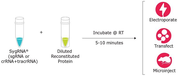
Figure 1.Cas9-GFP Protein GFP Complex
On-Target Activity
Other Cas9-GFP proteins on the market offer fluorescence, but with severely reduced cleavage efficiency (Figure 2). To solve this problem, our scientists designed and tested a panel of linkers and developed a proprietary configuration that provides the brightest and most potent Cas9-GFP nuclease to date.
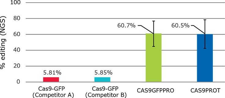
Figure 2.CAS9GFPPRO edits at rates 10X higher than other commercially available Cas9-GFP fusion products with no loss in activity when compared to non-GFP fusion proteins (average, n=10 different gene targets)
Off-target activity
RNP complexes naturally clear from the cell quickly to minimize potential off-target effects while point mutations in the chromosome-binding motif of SpCas9, as described by Slaymaker, et al., provide higher on-target fidelity than traditional Wild-type Streptococcus pyogenes Cas9. In addition, fusion of EGFP to the Cas9 protein is shown to have no effect on off-target activity.
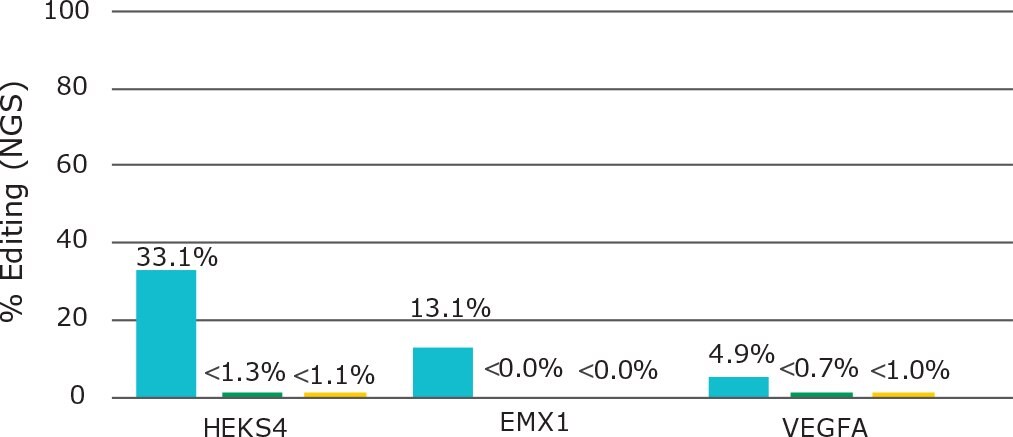
Figure 3.Enhanced specificity Cas9 (ESPCAS9PRO; yellow) and enhanced specificity Cas9-GFP (ECAS9GFPPR; green) have little or no detectable off-target cutting compared to competitor wild-type Cas9 protein (blue) at three different loci targeted with one part SygRNA™ synthetic sgRNA in HEK293T cells (n=3).
Flow Cytometry
The EGFP has an excitation peak at 488 nm, with an emission peak at 509 nm. This enables detection of the RNP complex in flow cytometers with standard GFP channels. Fusion of EGFP increases the utility of the protein, making it suitable for use in some flow cytometry, high content imaging and other screening applications.
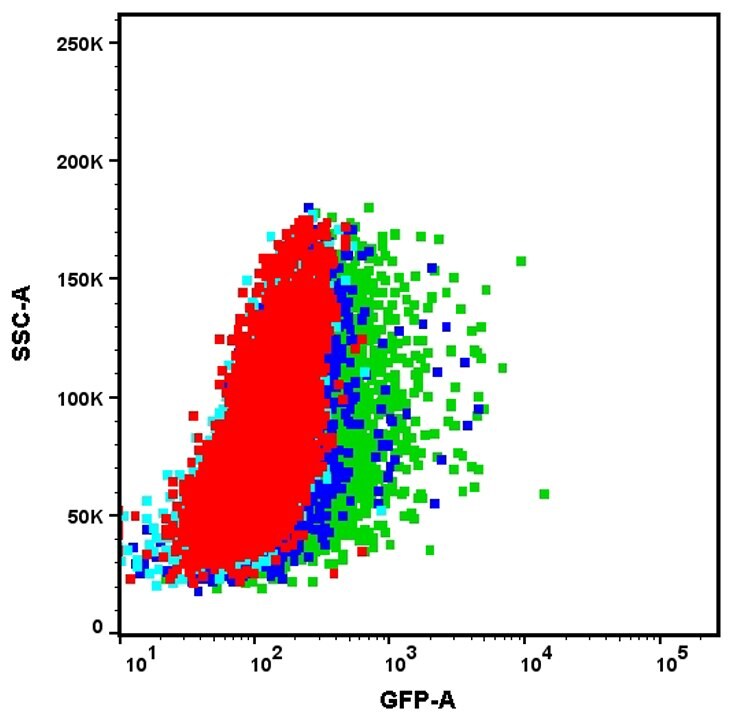
Figure 4.350k HEK293 cells were transfected with RNP complex containing 6 µg of Cas9-GFP protein and 5 µg of EMX1 one part SygRNA™ synthetic sgRNA (Cas9:RNA molar ratio 1:3). Within the cells, green fluorescence was analyzed via FACS at 24h, 48h, and 72h following transfection. By 72h, the GFP scatter has returned to the level of WT scatter, suggesting a complete clearance of the protein inside the cells
Fluorescence Microscopy
Under the microscope, Cas9-positive cells fluoresce green, verifying successful delivery. DAPI-staining of the same cells further confirms that Cas9-GFP is concentrated in the nuclei.
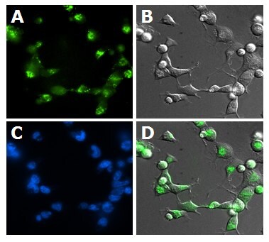
Figure 5.HEK-293T cells were nucleofected (Nucleofector™, Lonza) with a pre-complexed RNP composed of 150 pmol one-part SygRNA™ sgRNA targeting EMX1 and 50 pmol Cas9-GFP protein (CAS9GFPPRO-250UG) (3:1 gRNA:protein ratio). All images taken at 40x magnification approx. 24 hours after nucleofection. A) GFP B) Phase contrast C) DAPI D) Overlay of A and B.
To continue reading please sign in or create an account.
Don't Have An Account?